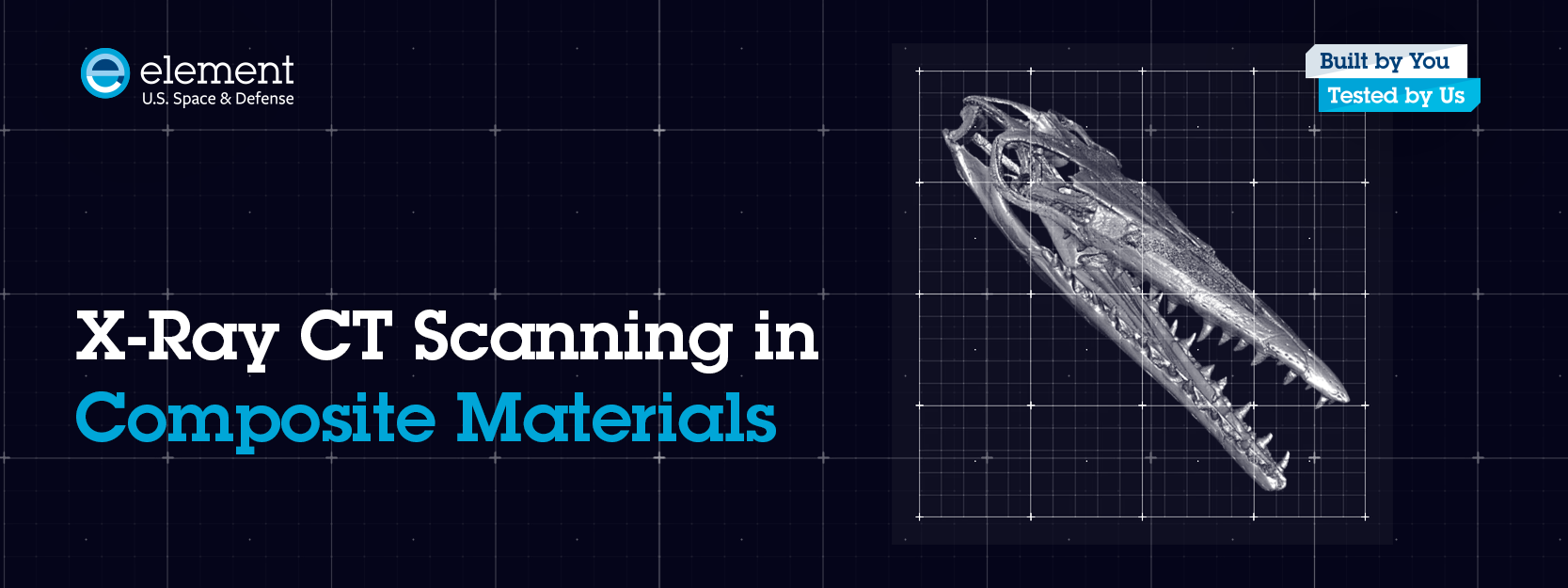
X-Ray Computed Tomography Scanning & Composite Materials
X-ray computed tomography (CT) scanning has become an essential technology for analyzing complex materials and structures with precision and depth. Originally developed for medical diagnostics, CT scanning has evolved dramatically over the past several decades to play a vital role in industrial and scientific applications, especially in the inspection of composite materials. Today’s advanced micro-focus CT scanning techniques enable researchers and engineers to visualize internal structures non-destructively, revealing details at micron and even sub-micron scales. Innovative facilities like Element U.S. Space & Defense’s Belcamp laboratory are pushing the boundaries of digital inspection, delivering unmatched insights into material integrity and performance.
The Evolution of X-Ray Computed Tomography Scanning Technology
X-ray inspection technology has advanced significantly since its early days. Introduced in the 1970s, X-ray computed tomography (CT scanning) has completely transformed medical diagnostics. By the 1980s, the emergence of micro-focus X-ray technology opened new possibilities in non-destructive testing for industrial and scientific applications. But it wasn’t until the early 2000s—thanks to major advances in X-ray detectors and computing power—that micro-focus X-ray CT scanning became commercially viable.
How Micro-Focus X-Ray CT Scanning Enhances Composite Material Analysis
With micro-focus CT scanning, data can be captured at incredibly high resolution, sometimes even at the sub-micron level. This makes CT scanning an extremely valuable tool in materials research, specifically when analyzing composite materials and their internal structures. The raw scan data, which is usually several gigabytes (20 GB+), can also be rendered in 3D and even numerically analyzed.
Visualizing Composite Structures: 3D Rendering & Fiber Segmentation
The image below shows a 3D rendering of a small section of a carbon-epoxy structure captured at approximately a 4-micron resolution. In this particular small composite block sample, the X-ray and imaging settings were optimized to enhance the contrast between the carbon fibers and epoxy resin. This enabled us to virtually segment and remove the resin material in order to expose the fiber structure. This data can be extremely valuable in evaluating structural properties of materials and different manufacturing processes. There are even software tools commercially available today that can numerically evaluate fiber consistency and orientation over an entire structure.
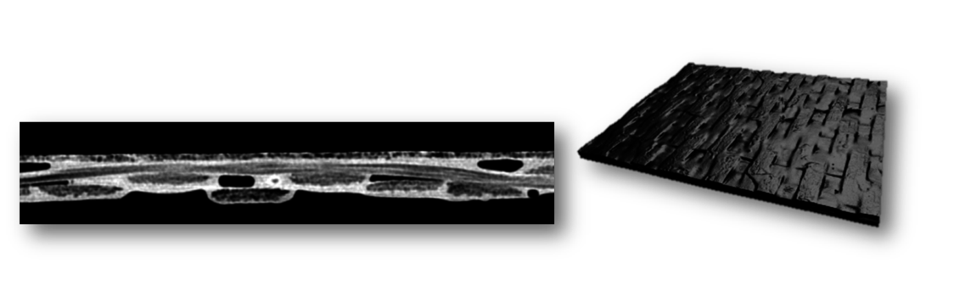
Analyzing Prepreg Composite Materials at Different Manufacturing Stages
X-ray CT scanning is a very versatile process that can be performed on many different materials and even at different stages of a manufacturing process. The images above, show a high-resolution CT scan of a prepreg composite that has not yet been fully cured. In the single cross-section image on the left, the brighter areas indicate uncured resin material, while small internal voids and openings are also clearly visible. These images can also be analyzed quantitatively to extract deeper insights—such as fiber volume fraction data—both at localized points and across larger regions.
Detecting Defects & Failure Modes with Micro-CT Scanning
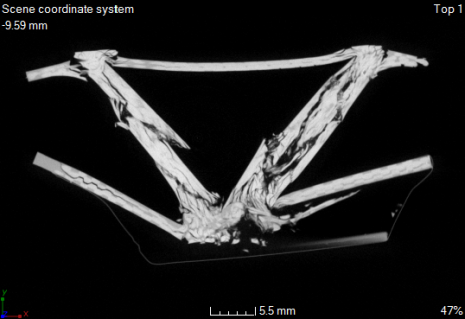
Even at the more “macro” scale, micro-CT is a powerful tool for structural and failure analysis. It allows small defects—such as porosity and thin delamination—to be visualized in high-resolution detail. Failure modes can be identified quickly, even within highly complex structures.
The image above shows a cross-section from a high-load bearing component that failed during mechanical load testing. Using micro-CT, both the origin and full extent of the failure can be examined without relying on destructive techniques that might compromise the sample or data.
X-ray CT scanning continues to expand its role in composite materials testing. It has proven especially effective in detecting previously hidden damage and failure mechanisms—while offering the added advantage of eliminating the need for often time-consuming destructive analysis.
Element U.S. Space & Defense: Innovating Composite Material Testing with Progressive XCT Scanning
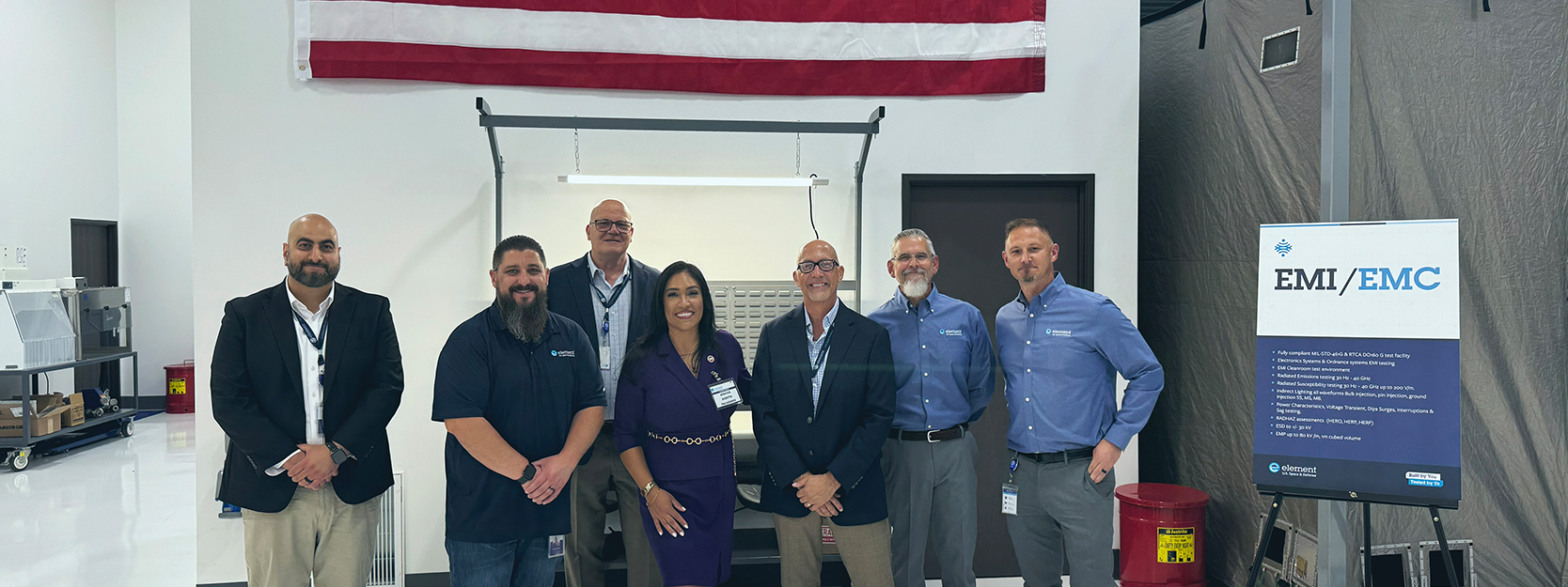
Element U.S. Space & Defense’s Belcamp, MD facility specializes in advanced digital X-ray and computed tomography (CT) imaging services, with a particular emphasis on Progressive XCT Scanning. Trusted by customers across a wide range of industries, the Belcamp team is comprised of specialists with deep expertise and training in high-energy and micro-focus applications. They consistently deliver accurate, timely results—even on the most complex projects. This capability enables Element U.S. Space & Defense to provide exceptional insights into component integrity and quality without compromising the part itself, further reinforcing its position as a leader in aerospace and defense testing solutions. To learn more about how our team can support your testing needs, reach out to our experts today.


Accessibility: If you are vision-impaired or have some other impairment covered by the Americans with Disabilities Act or a similar law, and you wish to discuss potential accommodations related to using this website, please contact us at (833) 673-8773